

Back to articles
Research & Diagnostics
3D Printing
Archaeology
Brain
Cognition
Dog
Domestication
Education
Endocast
Paleoanthropology
Skull
Training
Digital modeling of the dog’s brain to understand its domestication
MR
Mia Rozenbaum
10/28/2020
4 min read
0 comments
Share:
The endocranial cavity captures the imprint of the brain it encloses and can reveal many mysteries. Researchers are particularly interested in it to understand the morphological changes in the brain that led to the domestication of the dog.
An endocast—a cast of the endocranial cavity—is often used to study the external morphology of the brain (analysis of distance, surface area, or volume) without the physical presence of the brain itself. Particularly useful in paleoanthropology, this technique allows for the study of taxa of which only the skull remains.
Endocasts are generally made of latex or silicone, which presents certain drawbacks and can, in particular, damage bones that have become brittle over time. Neuroscientists, as well as paleontologists and medical personnel, now prefer to rely on structural imaging techniques.
Computed tomography (CT) is one of the most useful imaging techniques for digitizing bone structures and creating intracranial models. The resulting digital endocasts accurately reflect the morphology of the brain and even certain associated structures, such as the vascular system and cranial nerves. These digital casts can serve as the basis for creating detailed, high-quality three-dimensional (3D) models.
[caption id="attachment_9846" align="aligncenter" width="829"]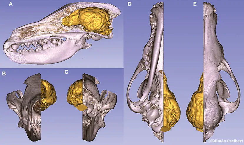 Volumetric skull model (with a median sagittal section) and the endocast of a gray wolf. (A) Left lateral view. (B) Rostral view. (C) Caudal view. (D) Ventral view. (E) Dorsal view.[/caption]
Just as reliable as latex or silicone endocasts, digital versions do not damage the original specimen, and most commercial CT analysis software is designed to perform all necessary calculations automatically. 3D printing makes it possible to physically produce and replicate these models infinitely at any desired scale, which can be useful in many fields, including archaeology and veterinary education.
A new study documents the methodology behind the creation of digitized endocasts
from canine skulls. Indeed, despite its relevance to both veterinarians and paleontologists, such literature was previously lacking.
The authors argue that comparing different endocasts could, in particular, help characterize the neuroanatomical variations associated with dog domestication. Indeed, behavioral changes linked to domestication may have an anatomical origin
and may have been caused by changes in size or proportions among different regions of the brain. This is especially true given that the mammalian brain—particularly its size—is influenced by natural selection and affects specific behavioral abilities.
[caption id="attachment_9845" align="aligncenter" width="893"]
Volumetric skull model (with a median sagittal section) and the endocast of a gray wolf. (A) Left lateral view. (B) Rostral view. (C) Caudal view. (D) Ventral view. (E) Dorsal view.[/caption]
Just as reliable as latex or silicone endocasts, digital versions do not damage the original specimen, and most commercial CT analysis software is designed to perform all necessary calculations automatically. 3D printing makes it possible to physically produce and replicate these models infinitely at any desired scale, which can be useful in many fields, including archaeology and veterinary education.
A new study documents the methodology behind the creation of digitized endocasts
from canine skulls. Indeed, despite its relevance to both veterinarians and paleontologists, such literature was previously lacking.
The authors argue that comparing different endocasts could, in particular, help characterize the neuroanatomical variations associated with dog domestication. Indeed, behavioral changes linked to domestication may have an anatomical origin
and may have been caused by changes in size or proportions among different regions of the brain. This is especially true given that the mammalian brain—particularly its size—is influenced by natural selection and affects specific behavioral abilities.
[caption id="attachment_9845" align="aligncenter" width="893"]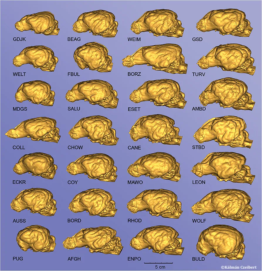 Endocasts of different canine specimens, showing the diversity of surface morphology. Left lateral view. GDJK, golden jackal; BEAG, Beagle; WEIM, Weimar Pointer; GSD, German Shepherd; WELT, Welsh Terrier; FBUL, French Bulldog; BORZ, Borzoi; TURV, Belgian Shepherd (Tervueren); MDGS, German Medium Spitz; SALU, Saluki; ESET, English Setter; AMBD, American Bulldog; COLL, Collie; CHOW, Chow Chow; CANNE, Cane Corso; STBD, Saint Bernard; ECKR, English Cocker Spaniel; COY, Coyote; MAWO, Maned Wolf; LEON, Leonberger; AUSS, Australian Shepherd; BORD, Border Collie; RHOD, Rhodesian Ridgeback; WOLF, Gray Wolf; PUG, Pug; AFGH, Afghan Hound; ENPO, English Pointer; BULD, English Bulldog. The endocasts are grouped into columns according to increasing total volume [thus, the endocast in the upper left (GJDK) represents the smallest endocast,[/caption]
To date, it remains unclear whether the relationship between brain volume and executive function reflects a large-scale evolutionary phenomenon or a unique consequence of primate brain evolution, which shows associations between brain volume and certain aspects of cognition.
The extraordinary degree of intraspecific morphological variation in domestic dogs offers a unique opportunity to study this phenomenon.
Endocasts of different canine specimens, showing the diversity of surface morphology. Left lateral view. GDJK, golden jackal; BEAG, Beagle; WEIM, Weimar Pointer; GSD, German Shepherd; WELT, Welsh Terrier; FBUL, French Bulldog; BORZ, Borzoi; TURV, Belgian Shepherd (Tervueren); MDGS, German Medium Spitz; SALU, Saluki; ESET, English Setter; AMBD, American Bulldog; COLL, Collie; CHOW, Chow Chow; CANNE, Cane Corso; STBD, Saint Bernard; ECKR, English Cocker Spaniel; COY, Coyote; MAWO, Maned Wolf; LEON, Leonberger; AUSS, Australian Shepherd; BORD, Border Collie; RHOD, Rhodesian Ridgeback; WOLF, Gray Wolf; PUG, Pug; AFGH, Afghan Hound; ENPO, English Pointer; BULD, English Bulldog. The endocasts are grouped into columns according to increasing total volume [thus, the endocast in the upper left (GJDK) represents the smallest endocast,[/caption]
To date, it remains unclear whether the relationship between brain volume and executive function reflects a large-scale evolutionary phenomenon or a unique consequence of primate brain evolution, which shows associations between brain volume and certain aspects of cognition.
The extraordinary degree of intraspecific morphological variation in domestic dogs offers a unique opportunity to study this phenomenon.
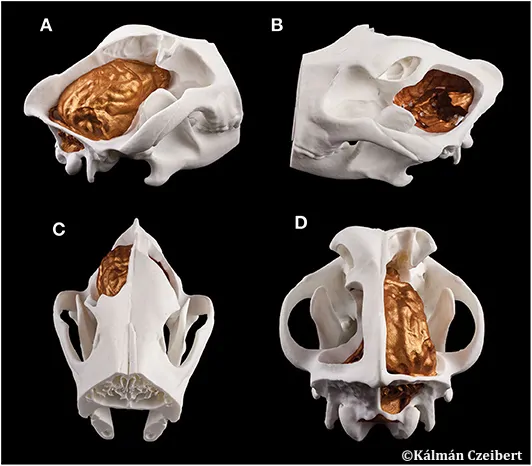 3D-printed composite model of a Weimar Pointer’s skull and endocast. (A) Right lateral view. (B) Left lateral view. (C) Rostrodorsal view. (D) Caudodorsal view.[/caption]
High-definition endocasts could help provide more detailed neuroanatomical measurements that could be used to more precisely link neuroanatomy to cognitive abilities, including social signals of domestication.By analyzing the relationship between the length and width of canine skulls and the surface areas of the rhinencephalic, prefrontal, and non-prefrontal cerebral convexity zones of the endocasts, the researchers were able to show that the ratio of the rhinencephalic area decreases with skull size. They hope that these results will serve as the basis for an analysis of skull and brain morphology, which will make it possible to link certain anatomical characteristics to behavior and cognition in dogs. Dogs could be considered a powerful model for studying the evolutionary links between cognition and neuroanatomy. Based on this principle, the study of detailed endocasts and neuroanatomical variations could provide a useful tool for identifying and documenting differences among breed groups. [caption id="attachment_9847" align="aligncenter" width="532"]
Read next
Research & Diagnostics
10/09/2026
WHO sets new diagnostic targets for Echinococcus granulosus in humans, dogs and sheep
Research & Diagnostics
10/09/2026
Nipah virus in wastewater: promising for early warning?
Research & Diagnostics
10/01/2026
Commentaires
No comments yet.
Sign in to comment